end tidal co2 range low
Capnography Waveform Interpretation Litfl Ccc Equipment Exhaled Carbon Monoxide End Tidal Co2 And Peripheral Oxygen Saturation Download Table End Tidal Co2 Emergency Medicine Icu Nursing Paramedic School Related. Ensure proper rate approximately 100min Ensure proper depth with adequate releaserecoil of thorax 12 thorax or minimum 25 inches Persistently low EtCO.

Utility Of Changes In End Tidal Carbon Dioxide After Volume Expansion To Assess Fluid Responsiveness In The Operating Room A Prospective Observational Study British Journal Of Anaesthesia
High quality CPR consistent waveform and end-tidal CO2 20 kPa.

. The number is called capnometry which is the partial pressure of CO 2 detected at the end of exhalation ranging between 35 - 45 mm Hg or 40 57 kPa. The waveform is called capnograph and shows how much CO 2 is present at each phase of the respiratory cycle. Definition of Low CO2 hypocapnia Hypocapnia hypocapnea also known as hypocarbia is defined as a deficiency of carbon dioxide in the arterial blood.
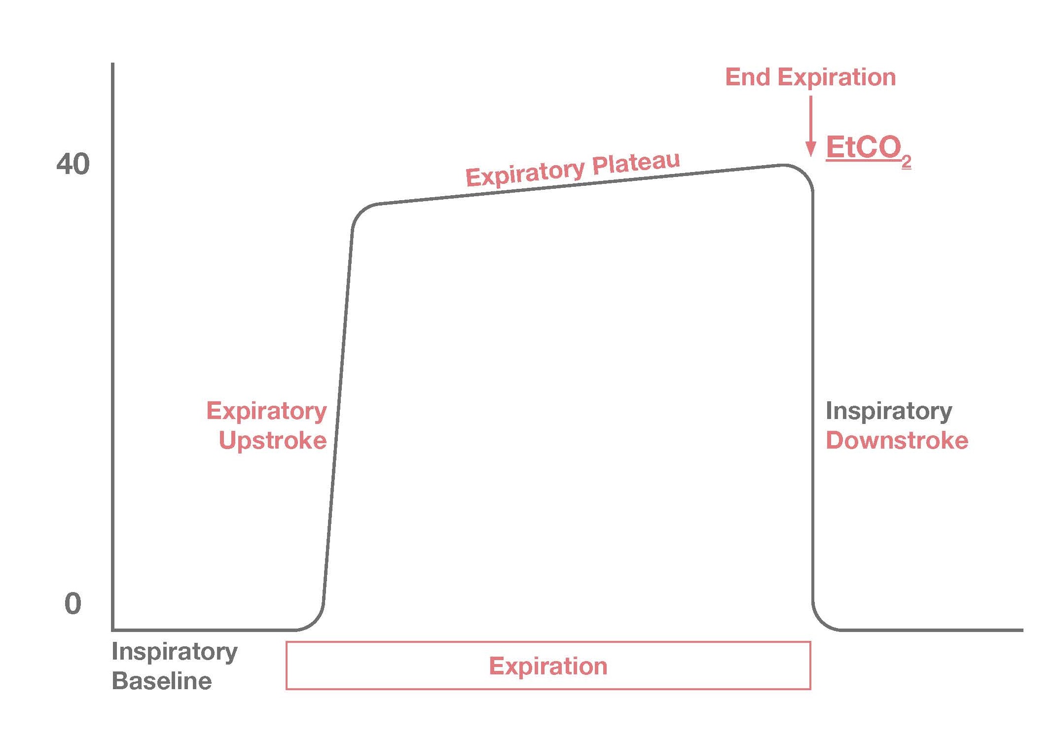
The normal end-tidal capnography wave form is basically a rounded rectangle. More recently a large group of medical doctors from several American hospitals tested over 100 patients and wrote an article End-tidal. The amount of CO2 at the end of exhalation or end-tidal CO2 ETCO2 is normally 35-45 mm HG.
From a retrospective single-center case-control study comparing low end-tidal carbon dioxide ETco2 postinduction in hypotensive patients due to anaphylaxis compared to other causes a low ETco2 contributed to the diagnosis of anaphylaxis. Waveform and end -tidal carbon dioxide EtCO2 values. This is a major respiratory symptom.
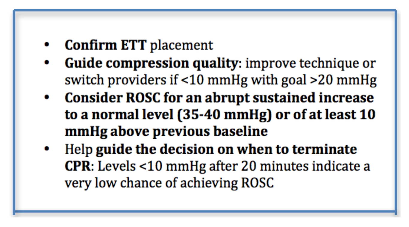
Measurement of a low ETCO 2 value 10 mmHg during CPR in an intubated patient suggests that the quality of chest compressions needs improvement. This is a major respiratory symptom. Misting increased SaO2 Types of End-Tidal CO2 Qualitative Yes or No.
Maximum end-tidal carbon dioxide Et co 2 within 5 minutes of the onset of mechanical ventilation in the operating room OR. End-tidal carbon dioxide cannot be used to rule out severe injury in. In addition a low P a CO2 level is correlated with increased risk of cerebral edema in children with DKA.
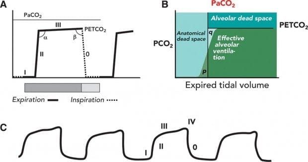
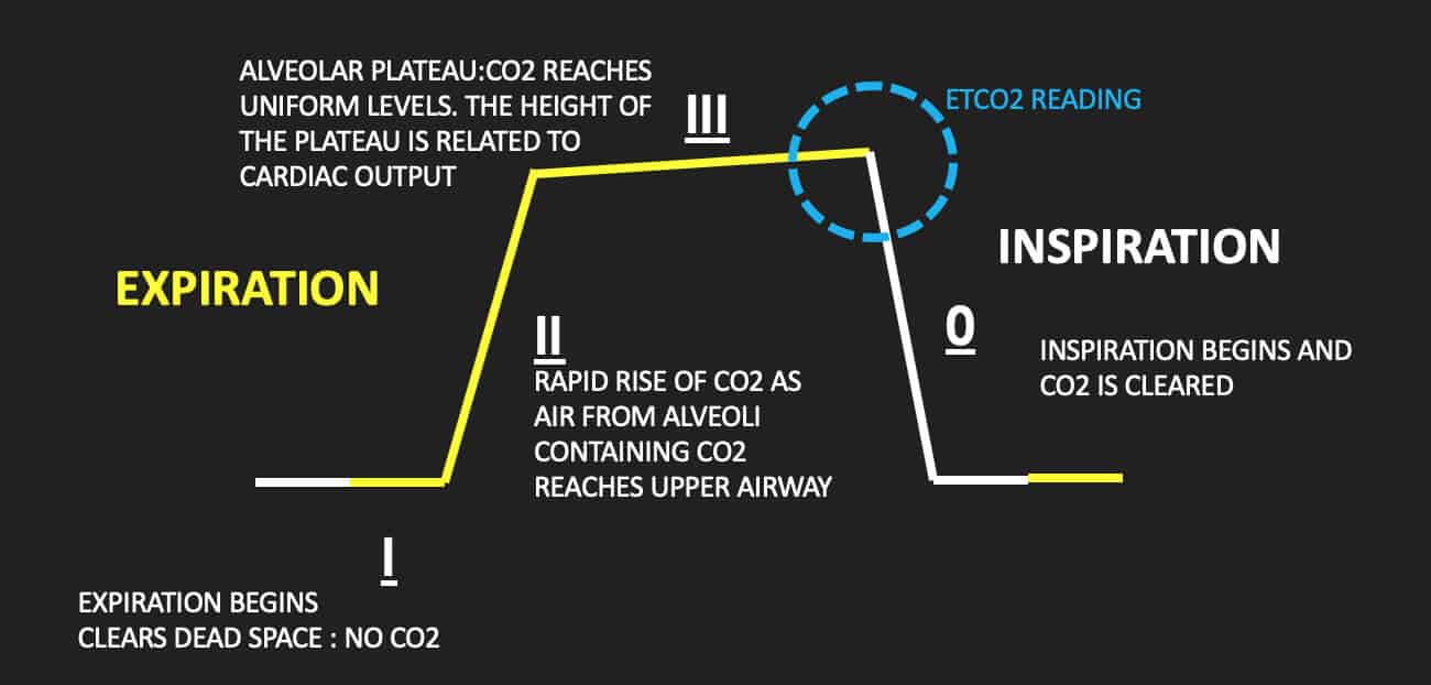
In addition the partial pressure of end-tidal CO2 PETCO2 during extremely low cardiac output has not been reported. 48 When a person is breathing out CO 2 the graph goes up. This effect could also be understood from the viewpoint of CO2 accumulation.
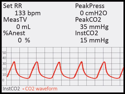
End tidal CO 2 monitoring is represented as a number and a graph on a monitor. When a person is breathing in it. Hence that persons capnography will show a very low end-tidal CO2 level but the alveolar and arterial CO2 pressures will be much higher and increasing in time.
The objective of the present study was to measure PETCO2 during well. The protocol prescribed to maintain ET co2 between 35 and 45 mmHg and to keep the systolic blood pressure SBP less than 180 mmHg before treatment and less than 220 mmHg after securing the aneurysm while maintaining a MAP greater than 80 mmHg. The results therefore suggest that in mechanically ventilated patients with severe postinduction hypotension ETco2 should be.
2 See Figure 1 p. If a person holds his breath that persons body accumulates CO2 and its etCO 2 is going to be zero. Most medical sources define hypocapnia as less than 35 mm Hg for partial CO2 pressure in the arterial blood.
Low end tidal co2 range Monday January 24 2022 Edit. End tidal normally 2-5 mmHg lower than arterial Comparing Arterial and End-tidal CO2 Review of Airway Confirmation Visualization Auscultation. Persistently low end-tidal CO2 check quality of compressions check ventilation volume if persistent may be a.
Sudden increase in end-tidal CO2 return of spontaneous circulation. The hinges represent the first and third quartiles the notches represent the 95 confidence interval CI of the median and the whiskers extend to 15 interquartile range. End tidal normally 2-5 mmHg lower than arterial Comparing Arterial and End-tidal CO2 Review of Airway Confirmation Visualization Auscultation.
The arterial CO2 value for normal breathing at rest is 40 mm. End-tidal clearance must be evaluated in the context of the patients perfusion status. A number of studies have shown that expired CO2 concentration is closely related to cardiac output but that cardiac output was not controlled as an independent variable.
Thirty-nine patients referred to our sleep laboratory because of suspected SAS and ten normal subjects were studied. The median ETCO 2 value was 32 mmHg IQR 27 38 mmHg range 18-80 mmHg. The range of values is unusually low.
End-tidal PCO2 abnormality and exercise limitation in patients with primary pulmonary hypertension. Negative Epigastric sounds Equal lung sounds Esophageal detector End tidal CO2 detector Secondary signs. Box plot with data points overlaid.
According to the book by Hockenberry and Wilson 2015 p 1140 normal values of ETCO2 are 30-43 mmHg which is slightly lower than arterial PaCO2 35-45mmHg. The researchers found that end-tidal CO2 could provide a highly sensitive predictor of return of spontaneous circulation during cardiopulmonary resuscitation MPR p791 Cantineau et al 1996. End tidal normally 2-5 mmHg lower than arterial Comparing Arterial and End-tidal CO2 Review of Airway Confirmation Visualization Auscultation.
ETCO2 emergency department monitoring and critical. This may result from such ventilatory problems as high mean airway pressure or inadequate exhalation time resulting in overdistention or from such circulatory problems as. Savastano S et al.
Dead-space ventilation results in ventilated alveoli with insufficient perfusion which leads to low ETco 2. Thus according to the relationship between ETCO2 and P a CO2. Forty-six of 96 48 95 confidence interval CI 38 58 patients had abnormal ETCO 2 values including 37 39 95 CI 29 49 with low ETCO 2 levels and.
Furthermore the directional changes of Petco2 during exercise and early recovery are in the opposite direction of normal. Chest compression provider tiring end-tidal CO2 value diminishes over time.
Capnography Waveform Interpretation Litfl Ccc Equipment
Schematic Diagram Of The Experimental Setup Etco2 End Tidal Co2 Download Scientific Diagram
Capnography Resus
3 Waveform Capnography Showing Changes In The End Tidal Carbon Dioxide Download Scientific Diagram

The Impact Of Ventilation Rate On End Tidal Carbon Dioxide Level During Manual Cardiopulmonary Resuscitation Resuscitation

Evaluation Of Suspected Pulmonary Embolism Utilizing End Tidal Co2 And D Dimer The American Journal Of Surgery
Capnoscan End Tidal Co2 Monitor Kent Scientific
Quantitative Waveform Capnography Acls Medical Training

3 Waveform Capnography Showing Changes In The End Tidal Carbon Dioxide Download Scientific Diagram

The Normal Capnograph Waveform Deranged Physiology

Exhaled Carbon Monoxide End Tidal Co2 And Peripheral Oxygen Saturation Download Table

The Capnometer Measurements End Tidal Co2 2009 Download Scientific Diagram
Emdocs Net Emergency Medicine Educationcapnography In The Ed Emdocs Net Emergency Medicine Education
2

Pre Hospital Capnography Ppt Download

Waveform Capnography In The Intubated Patient Emcrit Project

Waveform Capnography In The Intubated Patient Emcrit Project

Exhaled Carbon Monoxide End Tidal Co2 And Peripheral Oxygen Saturation Download Table

End Tidal Carbon Dioxide Recording Of Ventilated Children In Picu N 535 Download Scientific Diagram